RSS

Children and the Virtual Classroom

Corona has locked most of us up inside and along with us are the various addictive gadgets from the cell phones, computers, tablets, and televisions. The new norm is ‘work from home’ and employers are expecting more out of their employees because they are working in their comfortable homes! Some governments and educational institutions in their wisdom have also joined together to get students to also ‘work from home’ through unscientific online classes. I know of a school that has online classes for four hours a day, in two sessions of two continuous hours of lessons! What they don’t realize is that both the students and teachers are not tuned to such a sudden shift in the method of teaching.

To all of us, if not most of us, a classroom is a place where there is a one to one interaction between student and teacher. Most teachers are used to a room full of noisy, happy students whom they continuously monitor as a group. For the students, spending a day in school with their friends, sharing their lunches and playing together is a part of growing up and staying healthy. Unfortunately, the fear of Covid – 19 and the lockdown has turned the world upside down. The system of online classes, especially for school students here, is a stressful experience for not only the teacher and students but also for the parents and guardians of the child because the students are expected to be monitored during such online classes by a family member!

When you put a small child in front of a computer or a digital device there are certain health issues that we must take into consideration. It could be related to the device itself or the improper use of it. If you have a child attending online classes, keep the following things in mind.

The Device and its surroundings

Online classes are being conducted on either a computer, a mobile phone or tablet PC. Remember none of these gadgets can a substitute for a book. Neither can a keyboard, especially touch screen keypads, replace the pen and pencil.

Between the computer and the cell phone or tablet, the former is a better option because the screen is bigger. A page from a book can be read more comfortably on a computer than with a phone or tablet.  A normal A4 sized page can be viewed in the normal size on a larger computer monitor and may only require the page to be moved up and down. On a tablet or mobile screen you have to enlarge the page to see in a sufficiently large font size but then only a very small portion of the page can be viewed at any time and it will have to be scrolled in four different directions.

The computer can be kept on a table and viewed from a proper distance, but the smaller devices have to be handheld and so are too close to the eyes. If the online classes are going to be a long term and regular affair, it is better to get a desktop computer. These days basic computers are less expensive then mobile phones anyway! If not a tablet PC with a large screen is a possible option.

Most of these devices are designed with an adult in mind so setting up the computer for a child should be different. Normally, the screen should be positioned in such a way that we don’t have to bend our necks to look at it. It should be placed about two feet away and at a level where you only have to move your eyes slightly lower, about 15 to 20 degrees downward, to see the screen comfortably. For a child, you may have to make them sit on a higher chair so that the same angle of view is achieved. Similarly, the mouse and keyboard may also have to be repositioned to suit the child. The feet of very small children will not reach the floor while sitting on a regular chair, so a foot support would be a good idea. Alternatively, a table and chair that are lower and suits their height, could be a possible option.

All computers and digital devices are self-illuminated. Therefore, the ambient light in the room where it is used should neither be very dim nor too bright. The screen brightness should be adjusted to a level that is comfortable for the child and doesn’t make the child squeeze the eyes. Similarly, ensure that, especially if the device is near a window or light source, that the external light doesn’t hit the screen to cause uncomfortable glare.

Problems with excessive use of digital devices and computers

  1. Posture related problems. Most children using mobile devices are used to holding them close. They either sit hunched over it or lie stretched in an odd posture on a bed or settee. This can result in muscular aches, back and shoulder It is easier to control posture with a correctly adjusted desktop computer but with laptops or mobile devices it will require repeated reminders to maintain a healthy posture.
  2. Wrist pain. All keyboards and mice are designed for an adult so a very small child might find it very difficult to hold and manipulate the mouse or type comfortably. Excessive forcible twisting movements of the wrist can cause pain and eventually lead to a condition called carpal tunnel syndrome.
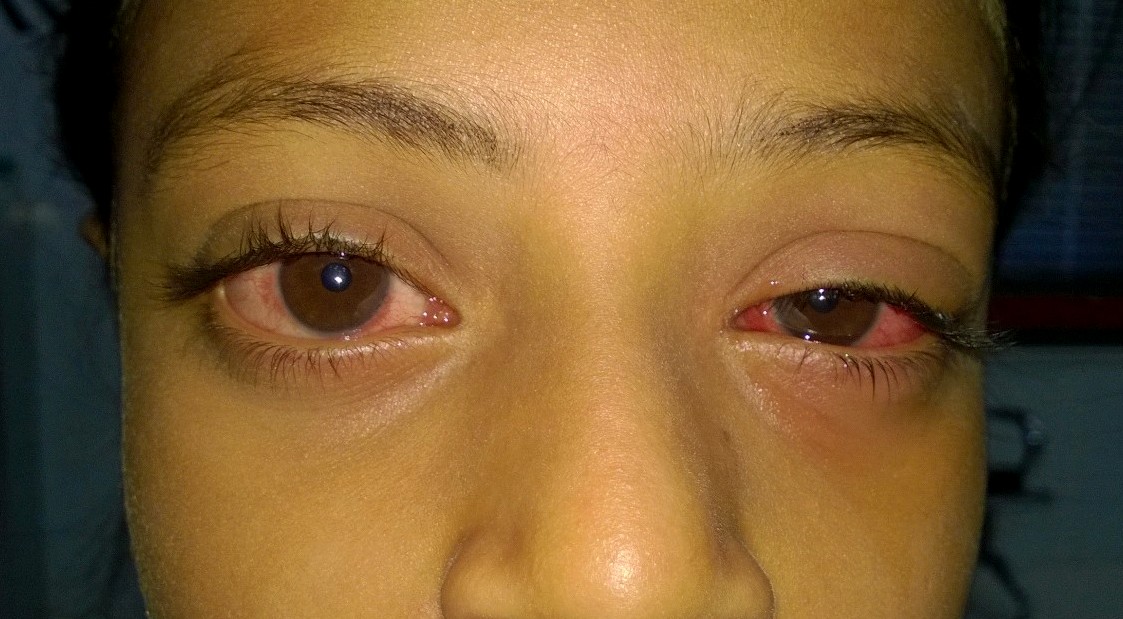
  3. Eye strain. Children cannot recognise the problems they experience while using these devices and will continue using them if they are performing and ‘enjoyable task’ like gaming. It is the responsibility of the elders to ensure that prolonged use should be curtailed with intermittent breaks. Otherwise, the children can end up with any of the following:
    1. Dry eyes due to staring unblinkingly at the screen for hours
    2. Focussing problem because of continuously looking at a fixed distance. The eyes have to be maintaining focus at a closer distance and the child eventually has ‘accommodative spasm’.
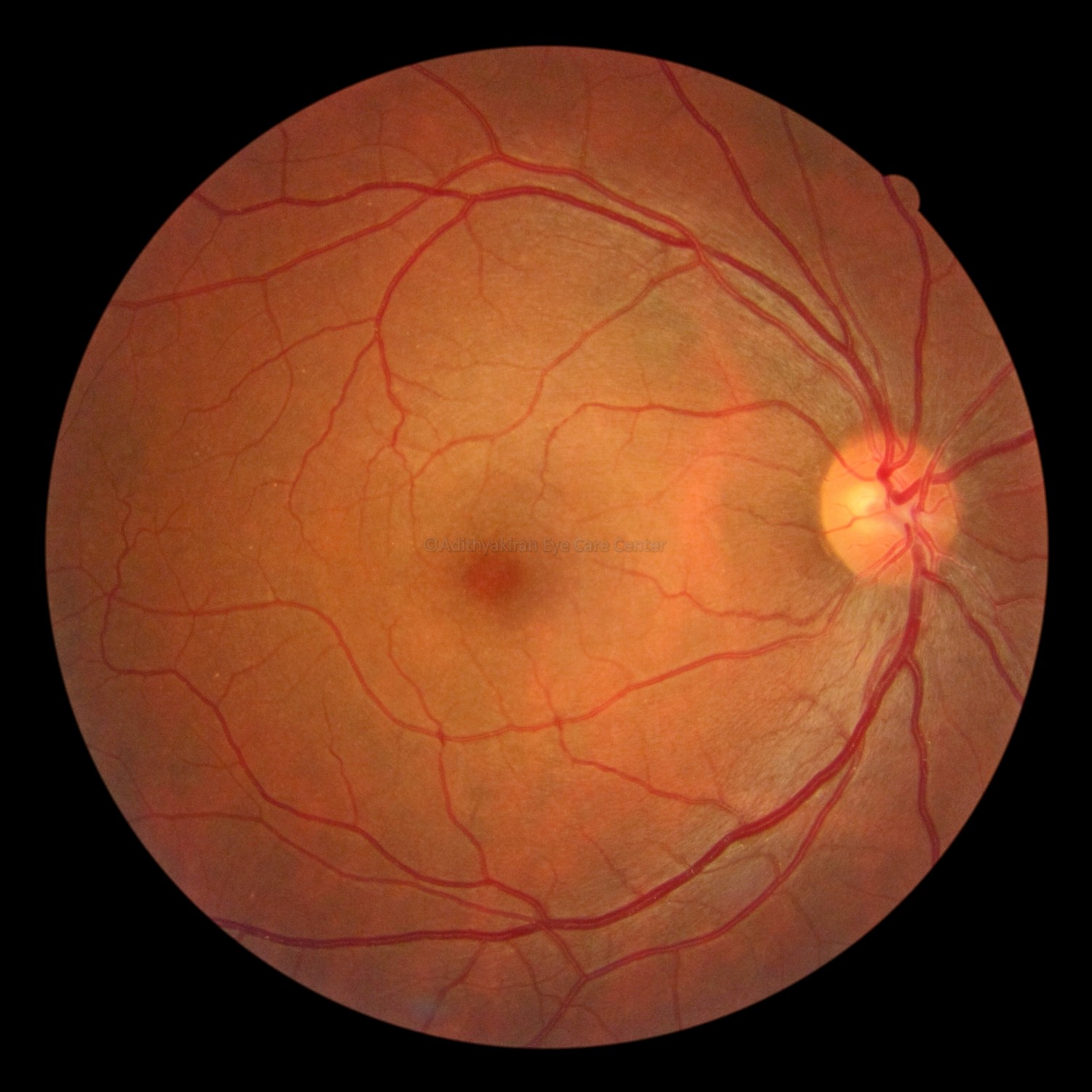
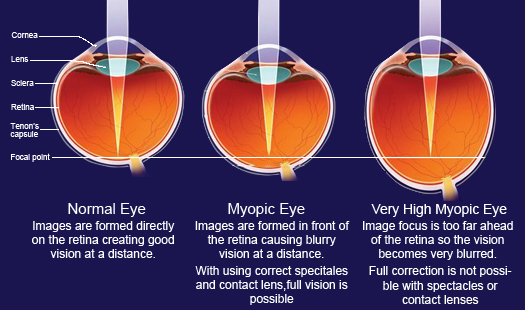
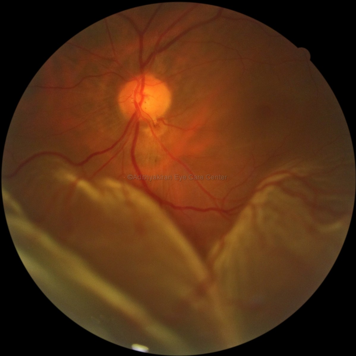
    3. Development of myopia (short sight) is common because the eye is always focussing at near objects and wont refocus for distance. In addition, not spending enough time outdoors can also contribute to progression on myopia.
  4. Children using these devices get fatigued easily because they are constantly sitting in one place and in an posture that doesn’t let the joints and muscles relax.
  5. A good night’s rest is essential for healthy life. Digital devices can affect sleep because of the ‘blue light’ effect if the child uses the device late at night.
  6. Children addicted to digital devices will show little interest in physical activity and that can cause obesity with its attendant risks.

Scheduling online classes appropriately

From the interaction I’ve had with the children, their parents and a few teachers, I feel there is something fundamentally wrong in the approach of the school authorities, politicians and other officials concerned with education. They seem to be in a hurry to make up for lost time and also ensure that running a school is a viable proposition in the middle of the this Covid crisis for which there is no end in sight.

Before planning classes we should look at how attentive a child will be in a monotonous environment.

A simple way of calculating the attention span of children according to child development experts is to multiply the age with a factor of two or three, (may differ depending on the source of information). This gives the number of minutes a child can pay constant attention to the task at hand. So if we calculate for the ages two to sixteen these are the approximate figures.

AGE IN YEARS

ATTENTION SPAN IN MINUTES AVERAGE ATTENTION SPAN

2

4 to 6 5

4

8 to 12

10

6

12 to 18 15
8 16 to 24

20

10

20 to 30 25
12 24 to 36

30

14 28 to 42

35

16 32 to 48

40


You can see that, younger the child, lesser the attention span. This calculation may be alright for a school in its proper environment, a classroom, but when it comes to online classes the arithmetic must change. In a classroom, there are classmates, friends and teachers interacting constantly on a personal level. To most children being in a noisy classroom is far more fun than interacting on an impersonal level with a device. The comforting touch of the teacher when things look down, exchanging childhood mischief with friends and sharing lunches are all missing! Instead, the child is forced to sit and concentrate at the job at hand, often with an elder supervising, in a rather stifling environment.

From the parent’s perspective, especially if they are both working, it is an exceedingly difficult proposition to manage this new system of online classes. Either one of them will have to take leave to remain with the child at home during these sessions. In case there are grandparents around, they could be the backup team but, in our environment, especially in poorer families they may not be tech savvy or educated enough to interact with the teachers on behalf of the children.

The problem does not end there. The teachers are also a suffering lot. One teacher told me that in the school she taught, the classes were conducted in two sessions. From 9 am to 1.30 pm for higher classes then from 4 pm to 7.30 pm for the smaller children. They would have a short break each hour but the difficulty was getting all the students back for the next round. They would have wandered off and will have excuses like internet issues and power failures to explain their absences! Not only that, the teachers would have time to prepare for the next day’s classes only after the day’s were over. Which means they would be up working till midnight and beyond!

We should take into consideration that even the teachers are probably more trained towards a classroom environment where they interact directly with their students. Therefore, when you are suddenly thrown into a situation where you are forced to learn and do something new and completely out of your comfort zone, problems are bound to crop up. 2020 as a year is going to continue in fits and jerks and we have to take each day as it comes. It is impossible to plan for more than a few days ahead as the behaviour of the pandemic is unpredictable. This year, not only for academics but everything else, will be a washout and therefore putting undue pressure on students and their teachers might backfire.

It is also a time where the parents will have to brush up on their digital skills or learn it from scratch. Online classes will be the norm for a few more months, perhaps longer, and only time will tell how beneficial it is to promoting education. Meanwhile, it would be better if parents, teachers and school managements are prepared properly for this unforeseen and unavoidable situation we are in.

Tips for managing an online class

  1. Set up the hardware in a properly lit room without any other distractions like televisions, refrigerators or snack cupboards. Children’s attention can easily be diverted.
  2. Make sure that the child is seated comfortably otherwise they will get restless and attention will wander.
  3. If an adult supervisor is in the room with the child, it is better they avoid fiddling with their cell phones or other gadgets. A newspaper or book is a better option.
  4. If there are other people in the house, ensure that they don’t disturb during the sessions. If they are watching TV (best avoided), listening to music or plain gossiping, keep the volume to the absolute minimum.
  5. Sitting in front of a digital device or computer forces the eyes to continue focussing at a very close distance, unlike in a classroom where the eye muscles can relax as the sight swings from the board to the desk and elsewhere. It means that the eyes become stressed faster so online classes should never run continuously for more than 20 minutes for small children or 30 minutes for bigger children.
  6. Remember the 20-20-20 rule. Continuous working on a self-illuminated screen at close distance can be tiresome, especially for a child. Therefore, after every 20 minutes (or 30 for older children) ask the child to look at some object 20 feet away for 20 seconds. That way, they eyes will be able to loosen up to relieve the strain of focusing continuously at the screen.
  7. Ask the child to blink frequently so that the tear film resurfacing can prevent drying up of the eyes. They will have to be reminded frequently till it becomes second nature to them.
  8. A break of 5 minutes should be given between classes, which will allow the child to relax focus and loosen up limbs and the body. If the teacher or parent can make the child do some simple loosening up exercise during the breaks, it would add to the excitement.
  9. Allow the child to take a few sips of water or juice in between. Snacks can be reserved for the longer breaks.
  10. It is better to confine the child to the room and not allow him or her to leave the immediate ‘classroom’ environment. Otherwise, they may lose the concentration. Let them learn to see the ‘virtual classroom’ like a real one and behave accordingly.

These are random thoughts that occurred to me as I’m seeing increasing numbers of children presenting with various complaints after the online classes have started. Please get in touch with me for any other doubts or clarification you may require.

Dr. Rajesh Radhakrishnan

info@adithyakiran.org

 
Leave a comment

Posted by on July 21, 2020 in Eye health


Tags: , , , , , , , ,

World Prematurity Day

I’m not an expert on premature baby eye but there is hope for these little one’s as this true story will tell.  It is the real life story of my friend and colleague’s journey with his son. If it touches a chord, please share it. There are many children like Ishaan who suffer from the want of proper evaluation in the right hands at the right time.

A Tale of Retonipathy of Prematurity

 
Leave a comment

Posted by on November 18, 2014 in Uncategorized


Red Eye & Your Child

A few days ago, I received a call in the morning. On the other end of the line was a worried voice. It belonged to an Obstetrician colleague of mine and she wanted to bring her one and a half year old grandchild to me because his eyes had been red for a week and was not responding to antibiotic drops she had given! I was a little taken aback. Here was a fellow professional asking me a health related question like a lay person, after using some drops in a one and a half year old child for a few days.

While there are many reasons for a ‘red eye’ in children two of the most common are the allergic conjunctivitis and viral conjunctivitis.

WP_20140224_008

Here is a list of common causes for red eye in children

  • Infectious conjunctivitis (bacterial or viral)
  • Allergic conjunctivitis (seasonal or perennial)
  • Conjunctivitis associated with dry eye
  • Lid margin infection (blepharitis or meibomianitis)
  • Foreign body
  • Injury to the surface of the eye
  • Lid margin deformities or misdirected eye lashes
  • Inflammation on the surface or inside the eye (episcleritis, scleritis and iritis)

This list is not complete but these are some of the causes for a child’s visit to an eye specialist.

In the part of the world where I live a common mode of transport in on scooters and motorcycles. In a country with a huge population and crowded roads two wheeler are a convenient way to avoid getting stuck in traffic.

OLYMPUS DIGITAL CAMERA

One unhealthy habit of most parents here exhibit is to load their entire family on a two wheeler without adequate protection.   OLYMPUS DIGITAL CAMERA

The smallest children are put on the foot board if it is a scooter……….. OLYMPUS DIGITAL CAMERA

………………or on the fuel tank if it is a motorcycle. OLYMPUS DIGITAL CAMERA

Only one in ten children wear any protective eye wear and more often, very grossly inadequate. Most of these children develop lid margin infection due to the exposure to the dust and dirt that gets into their eyes. That in turn along with the flow of air through the open eye causes an evaporation tear loss, the dry eye. Sometimes the dust itself can cause allergies.

OLYMPUS DIGITAL CAMERA

Red eye can present in  variety of ways and the probable cause can be ascertained from the presentation.

  • Mild redness could be due to dryness or allergy and usually resolves spontaneously if the exposure to the environment or allergen is avoided. A wrap around goggles and artificial tears are sufficient.
  • Redness that is mild to moderate associated with grittiness or itchy sensation is usually caused by dryness but early stages of viral conjunctivitis can also present similarly.
  • If the redness and itching are episodic or worsening during certain seasons it is generally due to allergy.
  • When the redness progresses over a few days and is confined to the junction of the cornea (the clear surface in the center of the eye) and conjunctiva (the white of the eye) it is usually vernal keratoconjunctivitis (VKC)
  • If the progression is rapid and generalized it indicates an infective conjunctivitis. Viral conjunctivitis is usually associated with itching and watering while bacterial conjunctivitis has a thicker greenish or greenish yellow discharge (pus).  It is also associated with swelling of the eyelids.
  • If redness is accompanied by pain and inability to open the eyes you have to suspect an injury or a foreign body on the surface of the eye.
  • Redness accompanied by worsening symptoms despite use of drops or ointments could be due to intolerance or allergy to the medicine. This is very common with over-the-counter medicines like neomycin and gentamycin.

Mild redness, especially due to allergy, responds well to artificial tears or anti-histaminic drops. In seasonal allergy a mast cell stabilizing medication maybe added, to be used through the season to prevent recurrence or worsening of symptoms. See a doctor if symptoms persist or worsen despite these simple measures. In severe cases steroid drops maybe indicated but they should be started only by your doctor.

Antibiotics have NO ROLE in allergic conjunctivitis. Most often when you approach the friendly next door pharmacy they give you an antibiotic or a steroid-antibiotic combination. By the time you go to your doctor the true picture if your original problem would have have been masked by these drops.

Similarly, in viral conjunctivitis too, antibiotics have no role. In fact using strong antibiotics like moxifloxacin or besifloxacin, as is becoming an unhealthy trend nowadays, can actually worsen your condition. You need anti-viral gels or ointments with some anti-histaminics or artificial tears to take care of the symptoms. Also remember, viral conjunctivitis will worsen symptomatically despite treatment over the first 5-7 days. It is s self limiting disease and usually goes away in 2-4 weeks. Your doctor might modify your treatment depending on the progression or improvement.

WHY MEDICINES DON’T WORK TO YOUR EXPECTATION

Most of the parents who bring in children with a red eye usually complain that the medicines given earlier did not work. This is because of the unreasonable expectations that parents have when their child is ill. All diseases have a natural course during which medicines have to be instilled as instructed by the doctor. Some conditions like seasonal allergic conjunctivitis will keep recurring till the child is in his or her late teens.

1. DON’T DELAY A VISIT TO THE DOCTOR. In my part of the world most children are brought in a few days after the problem has started. The stock excuse is that the parents expected the condition to resolve spontaneously “because the last time it went away in two days.” Remember, all red eyes are not caused due to the same condition. Only your doctor with special equipment can decide what is which.

2. DON’T EXPERIMENT. The other thing is, many parents tend to experiment because of their over-enthusiasm to see a quick cure. They use old drops that have been lying at home or some ‘grandma’s remedies’ with native medicines. This fact is never mentioned till they are questioned about it, but they will happily find fault with medicines given by their last doctor! If you have already used something, carry the earlier medicines so your doctor will know what you used.

3. DON’T OVERDOSE. Never over use the medicines. Diseases don’t get cured by overdosing. In fact you can cause more damage by using medicine above the recommended dosage. There are no special drops for very small children unlike what is available in oral or injectable forms. We have to make do with the same drops that are used for adults. Only your doctor can tell you the correct dosage and frequency. Using more medicine will not make the problem go away faster, especially conditions like viral conjunctivitis which have a natural course.

4. BE PATIENT. We doctors don’t use magic. We use medicines. Every medicine needs sufficient time to work and the course has to be completed. It your doctor has asked you to apply the medicine for a specific number of days and then go for a follow up, do so. Follow up is to see how much difference the medicine has made. If there are multiple drops don’t be in a hurry and instill them all together. There has to be a sufficient interval between drops to allow it’s absorption, usually 5-10 minutes. Longer the interval, the better. Another reason for poor response is because some children refuse to allow drops to be instilled in their eyes. How to overcome that problem is described below.

5.DON’T STOP MEDICINES WITHOUT INSTRUCTION. Antibiotics have different dosage schedules and courses. You have to use it for the prescribed number of days. Unauthorized stoppage or skipped doses can lead to resistance of infecting organisms to the medicine. If it is inconvenient to take a dose in the afternoon request your doctor to give a medicine that has a more convenient twice daily dosing. If you have been given steroids you should never stop it without being instructed to do so. They improve symptoms dramatically that you feel the problem is over. However, steroids have to be tapered off gradually. Abrupt stoppage can cause a rebound worsening of the condition being treated. Some drops may have to be used for months even if there are no symptoms. They are usually prophylactics.

6. DROPS v/s OINTMENTS. In very small children, instilling drops can sometimes be a real problem. Some children steadfastly refuse to open the eye and if you succeed in getting a drop instilled they squeeze their lids so hard that all the drops are driven out. In those situations an ointment or gel is a better option as they tend to ‘stick’ around long enough. Some children are very difficult to deal with, they will just not cooperate……

WP_20140224_005

……..they will kick and scream but will never open their eyes……..

WP_20140224_004

One option is to instill the medication while the child is sleeping but toddlers rarely sleep all day! For such children you will need three strong adults to achieve a successful instillation. One adult has to hold the arms at the elbows and keep it pressed against the side of the head firmly. Another has to hold the kicking legs at the knees, while the third person instills the medicine.

WP_20140224_006

Once it is done a few times the child also becomes cooperative and the eye improves dramatically! Then you have a happy child and an even happier parent! Don’t be scared to use a little force once in a while!

WP_20140227_005

PREVENT SPREADING OF RED EYE. Infectious conjunctivitis does not spread by looking. It spreads only by direct contact. If a parent instills the medication for a child, they should wash their hands with soap and water or use a hand sanitizer before touching anything else. All towels, sheets, pillow covers used by the child should be kept separately and washed separately. If the child is going to school a few days of avoiding classes will be beneficial for his or her friends and class mates.

NEVER SHARE MEDICATIONS. Drops or ointments prescribed for one child (or adult) should never be used by another child (or adult). The commonest reason for spreading of infectious conjunctivitis is shared medicines. Once a child recovers, collect all the used medicines and dispose them off properly.

Red eye in a child is by itself not always a serious condition. More often than not it is our impatience that leads to prolongation of the problem. Always follow instruction of your doctor. If things are worsening despite medicines go back and consult again with the same doctor. If you go to another doctor, the second one may not know how the problem was at the beginning. If things have improved, don’t stop medicines on your own. Go for a follow up as advised and stop the medicines only on the instruction of the doctors.

In red eyes caused due to allergy  ensure that you also avoid the allergen if it has been identified. Otherwise symptoms can recur every time the eyes are exposed to the allergen. If you are one of the people who uses a motorcycle or scooter, ensure that the child is seated behind you with adequate protection for the eyes. The best protection in my view is the swimming goggles. It will prevent even the most microscopic dust particle from entering your child’s eye and also prevent drying up of tears to to the air blowing through the eyes.

Treating red eye in children is a combination of medicines, preventive measures and protection.

 
1 Comment

Posted by on March 18, 2014 in Eye health, Uncategorized


Tags: , , , , , ,

Your Eye & Diabetes

November 14th in India is celebrated as Children’s Day because it happens to be the birthday of the first Prime Minister of independent India, Jawaharlal Nehru. In 1991, the International Diabetes Federation chose this day as the World Diabetes Day. It is a day for spreading awareness about diabetes and the threat it poses to the health of the sufferer, Since the disease is itself very silent and patients rarely feel very strong symptoms initially there is a casual attitude to the treatment advice give by health care practitioner’s, whatever their specialty.

chacha-nehru-wth-kids

Jawaharlal Nehru with children

One of the organs diabetes can affect is the eye. The ophthalmologist is placed in an enviable position because it is perhaps the only organ we can look into and actually see the changes diabetes has brought about. No other specialist has this unique advantage and they have to depend on indirect tests and investigation to decide if the concerned organs have been affected. It is no wonder that diabetologists insist that a patient with diabetes get their eye tested periodically. Therefore, it is only appropriate to understand how the eye can help you manage your diabetes.

WHAT IS DIABETES?

To understand how diabetes affects the eyes we should first know what diabetes is. Diabetes mellitus is a group of diseases characterized by high levels of blood sugar (glucose) that result from defects in insulin secretion, or its action, or both.  Insulin is a hormone produced by the pancreas and it lowers the blood glucose levels. When the blood glucose elevates (for example, after eating food), insulin is released from the pancreas to normalize the glucose level. In patients with diabetes, the absence or insufficient production of insulin results in elevation of blood glucose levels (hyperglycemia). Diabetes is a condition that can be controlled but not cured; therefore it is a condition that requires lifetime treatment.

There are two types of diabetes, Type 1 and Type 2. In Type 1 diabetes the onset is usually earlier, in individuals around 30 years and is a result of lack of production of insulin, therefore  treatment is primarily with insulin injections. Type 2 diabetes usually occurs in older adults. Here the pancreas does produce insulin but either the amount is insufficient or the body is unable to utilize it properly. This type can be controlled with tablets, exercise and lifestyle modification. Sometimes even Type 2 diabetics will need insulin.  South Asian populations, including Indians, are at a higher risk of developing diabetes than Europeans or White Americans. This is attributed to our genetic makeup, dietary habits, obesity and lack of exercise.

Over time, diabetes causes damage to various organs in the body and can lead to blindness, kidney failure and nerve damage. These diabetes complications are related to blood vessel diseases and are generally classified into small vessel disease, such as those involving the eyes, kidneys and nerves (microvascular disease), and large vessel disease involving the heart and blood vessels (macrovascular disease). Diabetes accelerates hardening of the arteries (atherosclerosis) of the larger blood vessels, leading to coronary heart disease (angina or heart attack) and strokes.

DIABETES AND THE EYE

Diabetes can affect the eye in many ways.

1.Temporary blurring

Fluctuations in blood glucose levels cause changes in the natural lens within the eye. The lens becomes stiffer and near focusing is affected so there may be an early onset of presbyopia. This can vary over time depending on the fluctuations in blood glucose levels. It is therefore a good idea to control your blood sugars before going to get your spectacles changed.

2.Cataract

High blood glucose levels can cause clouding of the lens of your eye, called cataract. Early cataract develops in the rear portion of the lens and though it may not cause a significant drop in you might experience glare and poor vision when ambient light is bright. Once cataract develops, the process is not reversible and the only option is surgery to replace the cataractous lens with an artificial lens.

3.Glaucoma

Though glaucoma is not a direct result of diabetes it has been found that there is a higher incidence of glaucoma among diabetics, up to 40% more, than in non-diabetics. The incidence increases with the duration of diabetes and age. Severe diabetic retinopathy can cause new blood vessels to grow into the front of the eye resulting in a rare form of glaucoma called neovascular glaucoma. It remains as one of the most difficult forms of glaucoma to treat. Surgery, in combination with laser can control the glaucoma for short durations but it is usually relentlessly progressive.

4.Diabetic retinopathy

The most serious effect diabetes has in your eye is on the retina, the thin membrane at the back of your eye that converts light from the objects we see into electrical impulses that are transmitted to the brain. A normal retina would appear very clean and uniformly shaded. The blood vessels travel from the optic disc in all directions and the macula, the part of the retina that controls your fine vision appears as a darker dimple in the center.

Normal Retina & Macula

Fundus picture of a normal retina & macula

Approximately 40% of persons with Type 1 and 20% of persons with Type 2 diabetes will develop retinopathy in long standing cases. Some studies claim that 50% of diabetics will develop some retinopathy after 10 years and 80% after 15 years, though this may not always be true.

NPDR with maculopathy

Diabetc retinopathy with maculopathy

Retinopathy is caused by diabetic damage to the small blood vessels inside the eye. The vessel walls become weak so they can bulge, leak or become blocked. Based on the changes seen they can be classified into two broad types, non-proliferative (or background) diabetic retinopathy and proliferative diabetic retinopathy.

4a. Non-proliferative diabetic retinopathy (NPDR)

This is the more common type of retinopathy and usually does not affect vision significantly. The patient may not, therefore, realize that they have a sight threatening condition. This condition is caused by the bulging (microaneurysm formation), bleeding (hemorrhages) or leaking of fluids (exudates) from the damaged normal retinal vessels.

Early NPDR1

Mild NPDR

Early NPDR2

Mild NPDR with exudates close to macula

If you can make out from the photos above, the macula is not involved very significantly. The vision is therefore not affected significantly. Once the macula is involved significantly, by either bleeding or exudation, there is a severe drop in vision (see photo below). This is called maculopathy (described later).

Maculopathy

Severe macular involvement

NPDR is sub-classified according to severity into mild, moderate and severe forms. Severe NPDR (see photo below) can also cause only a small drop in vision till macula gets involved. This type of retinopathy usually does not require any treatment except tight control of diabetes. However, if the macula is involved intervention becomes necessary.

Severe NPDR

Severe NPDR

Here is a video on diabetic retinopathy

4b. Proliferative diabetic retinopathy (PDR)

Proliferative diabetic retinopathy

PDR with new vessels over optic disc and elsewhere

When diabetes is uncontrolled retinopathy may progress from NPDR to PDR. Here, damaged blood vessels become blocked off starving the retina off oxygen. The body tries to compensate by encouraging new vessels to grow in place of the blocked ones. Unfortunately these vessels grow on the surface of the retina and into the vitreous (the jelly like substance within the eye). These new vessels are very weak and break easily leading to severe bleeding inside the eye (pre-retinal & vitreous hemorrhage). This bleeding results in sudden and profound visual loss. With time the blood can be reabsorbed

Traction retinal detachment

Retinal detachment due to traction from the dense scar tissue

In very severe case there is scarring wherever the new vessels grow (termed as neovascularisation) and shrinking of the scar results in traction and detachment of the retina (see photo above). At this stage, chance of visual recovery is almost impossible. Sometimes these vessels grow forwards into the front portion of the eye through the pupil and onto the iris. This results in a very severe form of glaucoma, the neovascular glaucoma.

4c. Diabetic Maculopathy

In case NPDR involves the central retina it is called maculopathy. The macula controls our fine vision and so any small damage in the form of bleeding or oedema (swelling of tissues) causes a severe drop in central vision. Though NPDR may not always need treatment macular oedema must be treated and good visual recovery is possible unlike in PDR.

MANAGING DIABETIC EYE DISEASE

The first priority in the treatment of diabetic eye changes is to control diabetes. No treatment is effective unless the cause of the disease is managed simultaneously. Fluctuation in vision usually stabilizes once the blood sugar levels are brought under tight control. Glaucoma can usually be managed with drops unless it is severe in which case surgical intervention maybe required.

Cataract, resulting from fluctuating sugar levels, can be prevented from progressing quickly if blood glucose levels are lowered. If there is no significant visual handicap surgery can be postponed. Hurrying cataract surgery in eyes with maculopathy may actually worsen the macular oedema. It is therefore better to treat the retinopathy before the cataract surgery.

NPDR without maculopathy responds well to tight control of diabetes. However, in many patients, especially with severe retinopathy some intervention might be required. Laser photocoagulation remains the mainstay of retinopathy treatment. In photocoagulation, tiny burns are made on the retina with laser. These laser burns seal the blood vessels and stop them from growing and leaking. Laser’s are applied in different ways depending on the severity of the retinopathy.

If there is significant new vessel growth (neovascularisation) large areas of the retina will need to be lasered. This is called pan-retinal photocoagulation (PRP), where hundreds of laser burns are made over two or more sittings. These burns help reduce new vessel growth by PRP reduces the risk of blindness from retinopathy, but it only works in conjunction with well controlled diabetes. This treatment is also used for neovascular glaucoma. Side effects of laser photocoagulation are usually minor. They include several days of blurred vision after each treatment and possible loss of side (peripheral) vision.

Click this link to see a video on PRP

Laser burns on peripheral retina (pic Courtest: http://www.nei.nih.gov/photo/keyword.asp?conditions=Diabetic)

Laser burns on peripheral retina (Pic Courtesy: http://www.nei.nih.gov/photo/keyword.asp?conditions=Diabetic)

Some people expect a dramatic improvement in vision after laser. That  will rarely happen because laser is only meant to limit the damage caused by diabetes. Since it actually ‘burns’ away the tissues there is an expected effect on the vision. When laser is applied extensively, peripheral retina loses some function. Besides the reduction in peripheral vision, night vision and colour vision is also affected.

In focal photocoagulation, the laser is aimed precisely at individual blood vessels that are leaking. This procedure is useful when these vessel are close to the macula, causing singificant macular oedema that results in reduced vision. Localized laser application is quicker and does not cause any effect on the vision. As a matter of fact the vision can improve as oedema resolves.

In the event where there is s bleeding inside the eye (vitreous hemorrhage) or there is a retinal detachment due to traction by the scared tissues inside the vitreous cavity, photocoagulation is no longer useful. The next option is vitrectomy, which is surgery to remove scar tissue and cloudy fluid from inside the eye. The earlier the operation occurs, the more likely it is to be successful. When the goal of the operation is to remove blood from the eye, it usually works. Reattaching a retina to the eye is much harder and works in only about half the cases.

Macular oedema

Macular oedema

When there is maculopathy with significant swelling of the retina, called clinically significant macular oedema (CSME), focal laser therapy can slow the leakage of fluid. Injection of some medications like anti-VEGF agents and  steroids into the eye can also help slow the growth of new blood vessels and reduce the leakage of fluid into the macula.

FOLLOW UP IN A DIABETIC PATIENT

If you have been diagnosed with diabetic retinopathy it is mandatory that you get your eyes examined regularly. Frequency of examination will depend on what type of retinopathy is present at the time of diagnosis. However, all follow up visits should include the following tests

  1. Visual acuity assessment
  2. Intraocular pressure measurement
  3. Dilated fundus examination
  4. Documentation of retinal findings by fundus photography
  5. Imaging of the retina to assess response to therapy

In patients with no retinopathy the recommended frequency is once in 2 years if diabetes is well controlled. If there is mild to moderate NPDR without maculopathy an annual eye check is adviced but if maculopathy is present an examination should be done every 6 months. In patients with severe NPDR and maculopathy an eye examination every 4 months is preferable. Patients with PDR will require follow up every 2-4 months depending on treatment adopted and presence of macular oedema.

During follow up besides the routine examinations few additional tests may be performed. Serial fundus photography will show the shifting pattern of the retinopathy before and after treatment. Sometimes an angiogram of the retinal will be done along with fundus photography to identify leaking points on the blood vessels so laser can be given effectively.

Serial fundus photographs

Serial fundus photographs

After laser photocoagulation the eye will be subjected to periodic retinal scanning with the Ocular Coherence Tomograph (OCT scan) to assess the regression of macular oedema. In a normal person the retina is a gently curving surface with a slight elevation in the posterior part(the macular area) . In the middle of this elevation is a small depression called the fovea.

3D view of a normal macula

3D view of a normal macula

On the OCT scan this depression can be clearly seen as a downward curve of the retinal surface.

Cross section of normal retina

Cross section of normal retina

In a person with diabetic maculopathy the foveal depression is sometimes filled up and bulges into the vitreous cavity. The exudates and fluid filled spaces can be seen in a cross section of the macula in such eyes.

Exudates and Oedema

OCT Exudates (white patch) and Oedema (dark area)

Level 3

OCT of Macular Oedema

3D image of diabetic retinopathy involving macula

3D image of diabetic retinopathy involving macula

Reducing the risk for retinopathy

Most diabetics will develop some amount of retinopathy eventually. After 15-20 years of diabetes every eye will show some signs of non-proliferative retinopathy with no significant effect on vision. This is because of certain risk factors that are beyond our control and listed below.

  • Age
  • Duration of diabetes
  • Ethnicity

However, there are risk factors that we can control and thereby reduce the chance of developing or worsening of retinopathy. They are:

  • Controlling of blood glucose levels
  • Control of blood pressure
  • Controlling cholesterol levels
  • Avoid smoking
  • Keeping fit and controlling weight

Things to remember about diabetes & your eyes

  1. Everyone with diabetes will eventually develop some changes in the eyes and this depends on the age and duration of disease.
  2. If your vision or eyes are not showing any symptoms it does not mean you have no eye problem. It is better to get your eyes checked before problems arise or vision deteriorates significantly.
  3. Discuss your eye problems with your physician who treats your diabetes, though they usually ask for an eye test report even before you have problems.
  4. Early eye problems can be managed by controlling your diabetes or by simple procedures like laser or drops.
  5. Don’t skip your annual eye examination because of the inconvenience of dilating. Your diabetic eye test cannot be completed without a dilated fundus examination.
  6. Monitor your blood sugar regularly. If you are using a home monitoring device, ensure you get your sugar levels checked once in a while in a laboratory also.
  7. Besides blood sugar level you also have to check your cholesterol, urea, creatinine and glycosylated hemoglobin (HbA1c) levels periodically.

If you or any of your family members are diabetic I hope you find this post useful. You may get in touch with me through email at either retina@adithyakiran.org or info@adithyakiran.org

 
Leave a comment

Posted by on November 11, 2013 in Uncategorized


Tags: , , , , , , , , , , , , ,

Diabetes and You

In November one disease that hits the spotlight is DIABETES.

The 14th of November is World Diabetes Day and there is a flurry of activity to spread awareness about this silent and deadly disease.

Diabetes & the Eye

Diabetes & the Eye

How much do you know about Diabetes? Take a quiz and see how your Eye-Q score is. If you are a diabetic you should score at least 9 out of 10!

Click here to take an online quiz from the National Eye Institute website 

You can also get more information on Diabetes here, the website of the International Diabetes Federation

 
Leave a comment

Posted by on November 11, 2013 in Eye health


Tags:

Bugs in your Eyes – 1: Lice

Do you know bugs can get into your eye or in the structures around it? This story is a little old. Happened a couple of years ago, in August 2011. I thought it deserves a separate post not only because the creature is interesting but also the issue highlights why doctors need to spend time with their patients.

This lady had gone to a doctor in a leading chain of eye hospitals for itchy eyes. She was given some drops for allergy and sent home. Since the complaint persisted the lady returned to her doctor who added a lubricating drop to the previous medicine. She was already on treatment for a skin condition with tablets (steroids and anti-histaminics) from many months. Finally, since she couldn’t get any relief she landed in my consulting room.

One look through my slit lamp my head reeled. There was a veritable zoo on her lid margin (click this line for the video link).

Phthiriasis palpebrum, actually pubic lice, were crawling on her lashes and some were borrowed into her lid margins. Many of the lashes had small nits attached to them. I did the normal treatment for the lice and sent her off with instruction to return after a week. When she returned a week later she was completely asymptomatic and her formerly, thickened lid margins were looking much healthier.

I asked her why she did not go back to her original doctor. She said, “He has no time to listen to my problem  or clear my doubts.” I had nothing to tell her after that.

The pubic lice, also called crab lice are usually found in the genital areas. They can be transmitted to the eyelid margins through hands, or other towels, bedding or clothing used by the infected individual. Is is also considered a sexually transmissible condition.

Treatment is done by paralyzing these creatures with pilocarpine and picking them off is an option. Sometimes all the eye lashes have to be trimmed to get rid of the nits adhering to them.

 
Leave a comment

Posted by on August 5, 2013 in Uncategorized

 
Link

Lists

Something everyone should be aware of when you go to visit your doctor. More often than not, many tests are done even though they give no additional information. Find out what is really required according to the problem you have with your health. Please pass this on to everyone you know who visit doctors regularly

(CLICK ON THE PICTURE or LINK ABOVE)

 
Leave a comment

Posted by on August 2, 2013 in Uncategorized


Tags: , , ,

Why do Doctors get Greedy? Ethics versus Money – Part 1

I know I’m going to raise a few eyebrows here and have a lot of folks in my profession fuming at this bluntness. when I told a colleague about my intention I was told that I would be branded a black sheep and a traitor. That’s Ok. I’ll live with ‘branding’, because when I took the Hippocratic Oath in Medical College it was with the intention to serve my patients, not manufacturers of equipment,  drugs or implants that I use. Unfortunately, a lot of my colleagues tend to forget that we became health professionals to serve the patients who come to us so trustingly, putting their faith and hard earned money in front of us to make them better.

greedy-doctors41

Doctors getting greedy are not a new phenomenon but the immediate stimulus for me to drop the thread on Glaucoma temporarily was this article. (Click the line in red below to go to the page. You may have to sign in to read the article)

US Cardiology Scam on MEDSCAPE

When a patient goes to a doctor with a problem he or she goes with the hope that their problem will find a solution in the doctor’s office. The choice of the physician depends on the faith the patient has in that particular doctor. Sometimes, most often, it is influenced by the reputation of the doctor in the community and how he or she handles their patients. These days, when health care is slowly getting converted to a booming business, another factor has crept in, GADGETRY.

There are many patients who believe that a doctor is only as good as the type and number of gadgets he uses in the clinic. These type of patients are the ones that really are responsible for the gradual deterioration of healthcare standards. Doctors despite all their specialized training become glorified technicians. Clinical acumen gets replaced by instrumental dependance because the patient is unhappy if the doctor does not examine him or her with more than a few fancy gadgets!

Most corporate hospitals in my part of the world have something called the Master Health Check or Executive Health Check. If you are a person with no health problem this is the best way to ‘become a patient’! There is a series of tests performed, investigations done, scans taken and specialists consulted. At the end of it we are disappointed to find there is something ‘slightly wrong’ in one of the reports. So further tests or detailed investigations are done, that are chargeable separately (not included in the package deal!). Finally, you end up with a fat bill and a big list of, mostly unnecessary, medication with instructions to repeat tests and medication regularly! Someone who went in to confirm that they are hale and healthy steps out of the hospital, branded a patient.

What most of us forget is that as we age some parts of our body also wear down and stop working as efficiently as they did when we were younger. Arteries clog, skin and muscles lose their tone, and bones become less dense. It is a normal part of aging. If you end up doing one of these detailed checks on your body you are bound to find a lot of things that are a little off from the so called normal values. Even normal values are different, from lab to lab, time to time and person to person. You don’t have to be alarmed if they are not significantly different from the prescribed normal limits.

We love to be a ‘patient’ and pop pills even though many are unnecessary. Many of us beyond 50 years take an cholesterol lowering agent, a blood thinning agent, a set of vitamins and anti-oxidants and an antacid to prevent these totally unnecessary drugs from ruining our gut. All, with a  fervent hope that we will not die of heart attack or cancer! No wonder pharmaceutical firms are the best business to invest your money in. After all population of old hypochondriacs is booming.

The following links will take you to two interesting articles related to this topic that I have just started.

Strategies to Avoid 5 Highly Overused Treatments

‘Choosing Wisely’ targets 90 more dubious tests, therapies

 
1 Comment

Posted by on August 2, 2013 in Uncategorized


Tags: , , , , , ,

What is ‘proper’ eye testing for a myopic person?

As usual this interjection is because of a patient who walked in just now. She is a spectacle wearer since the age of 5. She has been having little annoying dots in front of her eyes that she couldn’t get rid off. I was querying her about her history of using spectacles and the conversation went something like this.

Me: When did you get your spectacles last?

She: A year ago.

Me: Where did you get your eyes checked then?

She: At the optical shop. (I’m not mentioning the name since it is close to my clinic).

Me: Who checked your eye there?

She: There was a doctor there.

Me: Were you dilated and examined.

She: No. They tested me on a computer.

Me: Then where did you get your glasses prescribed from before that?

She: At XXX hospital, a year before that.

Me: Were you dilated then?

She: No.

Me: Then, when was the last time you got dilated?

She: Don’t remember

I stopped asking her anything further. What I’m trying to highlight here is that in our very hurried lives we forget to take care of one of God’s most precious gifts. Our eyes. Since our lives are tightly bound by the wrist watch that we all wear, it has become difficult to go to an ophthalmologist to get our eyes checked properly. After all who’d want to sit for an hour with uncomfortable dilating drops put into your eye till you are completely unable to see clearly. What’s more annoying is that the effect of the dilating drops last the rest of the day, so that ruins your chances of watching that interesting movie on TV!

Unfortunately, as ophthalmologists, our job is not done by just prescribing your spectacles. We have to look at your eyes outside to inside, and ensure that you are not at risk for any complications of myopia. Most of the complications are related to the difference in the structure of the myopic eye.

The Myopic eye is essentially a ‘stretched’ eye. Which means, the eye is longer from front to back when compared to a normal eye. This mean rays of light have to travel further backwards to fall on the retina. Unfortunately, only the outer layers of the eye stretch; the other structures in between do not stretch or change proportionately. Therefore the crystalline lens fails to bring light entering the eye to a point focus on the retina. This focal point, at which a clear image is obtained falls somewhere in front of the retina.Myopia

When myopia is mild or moderate good vision can be obtained with corrective spectacles. However, even such eyes can develop some changes in it that can cause severe vision loss. The belief that everything is alright with your eye once your spectacle number stabilizes is wrong. There are changes that take place within the eye, that you may not be able to notice, but which can cause some serious problems with vision.

These changes are related to the stretching of the eyeball due to progression if myopia. Most of the vision threatening changes occur in the far periphery initially. The retina is stretched so much that it starts ‘cracking‘, something similar to old clothes getting frayed after many washes.

Pic courtesy: Optos

Pic courtesy: Optos

This ‘cracking’ is called lattice degeneration (see above). These cracks can eventually stretch too much to form ‘tears’ or ‘holes’. (see below)

vodvos-retinal-hole

Once a hole or tear appears you might get symptoms of streaks of light flashing past your face. These flashes can be normal too. The next time you feel a sneeze coming up just close your eyes and let go. You’ll see white spots of light flying on the periphery of your vision that will last for a few seconds.

Sometimes, especially in higher degrees of myopia the vitreous (the jelly like substance inside the eye) liquifies and it is this liquified vitreous that appears as small dots or shadows (called floaters) in your vision when you look at a bright background. If this fluid vitreous goes though the tear it can separate the retina from the underlying layers, a condition called retinal detachment. (see below)

RD

There will be a sudden drop in vision with flashes that are continuous. This is an ophthalmic emergency. Unless the retina is surgically replaced immediately there is great risk of permanent loss of vision. Retinal detachment can be prevented to a large extent by using lasers to ‘fence off’ the hole (see below) and prevent the liquid vitreous going under the retina.

lasered

In some advanced cases of myopia the eyeball stretches backwards so much that the retina and underlying structures get thinned and damaged near the optic nerve head and the macula (the part of the eye that controls fine vision). That condition is called a posterior staphyloma and it usually results in poor central vision.

early staphyloma Pathological myopia

Compare this to a relatively normal fundus in this picture below.The disc and the blood vessels are spread out on a very uniform background retina unlike in the pictures above.

normal fundus

So the next time you go to your ophthalmologist don’t just say, “I’ve come to check my number”; say “I’m here to check my eyes inside and out”.

Make sure you get all these tests done:

  1. Visual acuity with and without spectacles
  2. Best Corrected Visual Acuity if your spectacles feel inadequate
  3. Intraocular pressure
  4. Retiniscopy
  5. Ophthalmoscopy, including indirect ophthalmoscopy if you have higher degrees of myopia

EVENING UPDATE 6.30PM

I’m updating here because of a patient who walked in an hour ago. She had undergone LASIK in 2002 and her spectacle number before the procedure was -14 diopters. After LASIK it had reached near about zero and is still stable. If you have undergone LASIK to get rid of your glasses please do keep in mind that this procedure only alters the front of your eyes. All the serious changes occur within the eye and therefore you need to continue getting your eyes checked every year, as described above. Otherwise, subtle progress can be missed and you could end up with a sight threatening complication!

 
2 Comments

Posted by on June 20, 2013 in Uncategorized


Tags: , , , , , , , , , , , , ,

Glaucoma (Part 3) – “Confirming the presence of the Silent Thief”

In the previous part I had told you how to identify if you are a potential candidate for developing Glaucoma, the Silent Thief of sight. Now it is time to look at the different ways you can confirm if you have the disease.

Detecting glaucoma, in most instances, does not require sophisticated equipment. Ophthalmologists can identify features of the disease by just using some common instruments that they already possess. If a systematic approach is done the disease can be picked up early.

1. SLIT LAMP EXAMINATION

Slit Lamp

During preliminary examination with the instrument called the slit lamp the ophthalmologist may see some features that point to the presence of glaucoma. There maybe shallowing of the anterior chamber, the space between the cornea and the iris suggesting a possibility of angle closure glaucoma. There can be pigments on the inner surface of the cornea, or some flaky white material on the surface of the lens called pseudo-exfoliation.

2. TONOMETRY

The next step will be to see if your intraocular pressure (IOP) is raised. Remember though, glaucoma needn’t always be associated with an elevated IOP. There are eyes that have glaucoma even with normal or low pressures. IOP, therefore, is not the main criteria for deciding if you have glaucoma. IOP is measured by an instrument called the applanation tonometer, attached to the slit lamp. After instilling a drop of anesthetic the tip of the instrument is made to touch the dome of your cornea and a reading is taken from a rotating dial on the side of the instrument. There are other instruments to measure the IOP but the applanation tonometer is take as standard.

GAT

3.PACHYMETRY

Sometimes the IOP maybe normal or below normal but other features may give an indication that the eye has been affected significantly by glaucoma. The ophthalmologist may then do a pachymetry, a test done to measure the thickness of the cornea.

Pachymeter


It is not unusual to have people with myopia (short sightedness) to have a thin walled eye. This gives a falsely low reading of IOP. The reverse maybe true in other eyes. Due to a thicker cornea, the recorded IOP might by high, but there may not be any evidence to suggest glaucoma. Such eyes with higher IOP but without features of glaucoma are designated as Ocular Hypertension, and will need follow up to see if they progress to glaucoma.

4. OPTIC NERVE HEAD EVALUATION

After having measured the IOP the next step is to look inside your eye to see if there is any physical damage to your optic nerve head (ONH). This is the most important part of the examination that lets the ophthalmologist decide if your eye has been affected by glaucoma and if you need any treatment. The ONH can be examined by a simple hand held instrument called the ophthalmoscope; a self illuminating, hand held instrument that is held close to your eye and the doctor looks inside from the other side.

ophthalmoscope

For greater details, a small, powerful hand held lens (a 78 or 90 diopter) is used in combination with the slit lamp bio-microscope. It gives a bigger 3D view of the ONH and any damage can be assessed easily.

90D lens

This is the most important step in the assessment of a person suspected to have glaucoma. Features onthe ONH or surrounding retina can instantly clinch the diagnosis of glaucoma. The doctor will look for certain changes in the ONH that will show if glaucoma has already damaged the eye. I will dwell a little longer on this part of the exemination because, of all the examinations, this is the single most critical test that gives the doctor a hint that you have glaucoma.

Normal optic disc

To know what features suggest damage to the doctor, you should know what a normal ONH looks like. The optic nerve is the bundle of nerve fibres that start from your retina and go towards the visual cortex of your brain. As they exit the eye they form a distinct round or slightly oval disc, orange in colour, called the optic disc (see figure above). In the center of the disc (blue circle in figure below) is a smaller, light yellow coloured portion called the optic cup (yellow circle in figure below). This cup is usually well centered or slightly eccentric, when surrounded by heathly nerve fibers.

Disc & cup


The portion between the blue and yellow ring represents healthy nerve fibers and they are present in all quadrants of the disc in slightly varying widths. This part is called the neuro-retinal rim (NRR). The first sign of damage is a narrowing of the NRR, (red arrow) sometimes all the way upto the edge of the disc.

Rim narrowing

Sometimes associated with this narrowed rim there can be other signs that indicate the presence of active glaucoma. There might be small hemorrhages (bleeding) along the disc margin (yellow arrows in the picture below).

Hemorrhages

In some eyes, there is a dark band on the retina corresponding to the area of narrowing of NRR (between yellow arrows in the picture below). They are areas of retinal thinning due to loss of some tissue there. They are called retinal nerve fiber layer (RNFL) defects. They usually correspond to defects in the visual field tests.

RNFL defect

You can see that an ophthalmologist can make an instantaneous diagnosis of glaucoma if any of the above mentioned findings are there in a patient’s eye. Very simple instruments and equipment are need to make a diagnosis. It is only in cases where the findings are borderline that we need sophisticated instruments to give additional information to support the diagnosis.

Once glaucoma is detected the next step is to assess how much damage it has done to the eye. Damage to visual function usually occurs from the periphery of your vision. This is assessed by a test that plots the visual field. More about that in the next post. 

 
Leave a comment

Posted by on May 30, 2013 in Eye health


Tags: , , , , , , , , , , , , , , ,