RSS

Tag Archives: indirect ophthalmoscopy

What is ‘proper’ eye testing for a myopic person?

As usual this interjection is because of a patient who walked in just now. She is a spectacle wearer since the age of 5. She has been having little annoying dots in front of her eyes that she couldn’t get rid off. I was querying her about her history of using spectacles and the conversation went something like this.

Me: When did you get your spectacles last?

She: A year ago.

Me: Where did you get your eyes checked then?

She: At the optical shop. (I’m not mentioning the name since it is close to my clinic).

Me: Who checked your eye there?

She: There was a doctor there.

Me: Were you dilated and examined.

She: No. They tested me on a computer.

Me: Then where did you get your glasses prescribed from before that?

She: At XXX hospital, a year before that.

Me: Were you dilated then?

She: No.

Me: Then, when was the last time you got dilated?

She: Don’t remember

I stopped asking her anything further. What I’m trying to highlight here is that in our very hurried lives we forget to take care of one of God’s most precious gifts. Our eyes. Since our lives are tightly bound by the wrist watch that we all wear, it has become difficult to go to an ophthalmologist to get our eyes checked properly. After all who’d want to sit for an hour with uncomfortable dilating drops put into your eye till you are completely unable to see clearly. What’s more annoying is that the effect of the dilating drops last the rest of the day, so that ruins your chances of watching that interesting movie on TV!

Unfortunately, as ophthalmologists, our job is not done by just prescribing your spectacles. We have to look at your eyes outside to inside, and ensure that you are not at risk for any complications of myopia. Most of the complications are related to the difference in the structure of the myopic eye.

The Myopic eye is essentially a ‘stretched’ eye. Which means, the eye is longer from front to back when compared to a normal eye. This mean rays of light have to travel further backwards to fall on the retina. Unfortunately, only the outer layers of the eye stretch; the other structures in between do not stretch or change proportionately. Therefore the crystalline lens fails to bring light entering the eye to a point focus on the retina. This focal point, at which a clear image is obtained falls somewhere in front of the retina.Myopia

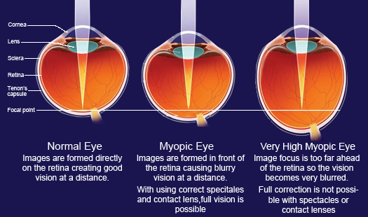
When myopia is mild or moderate good vision can be obtained with corrective spectacles. However, even such eyes can develop some changes in it that can cause severe vision loss. The belief that everything is alright with your eye once your spectacle number stabilizes is wrong. There are changes that take place within the eye, that you may not be able to notice, but which can cause some serious problems with vision.

These changes are related to the stretching of the eyeball due to progression if myopia. Most of the vision threatening changes occur in the far periphery initially. The retina is stretched so much that it starts ‘cracking‘, something similar to old clothes getting frayed after many washes.

Pic courtesy: Optos

Pic courtesy: Optos

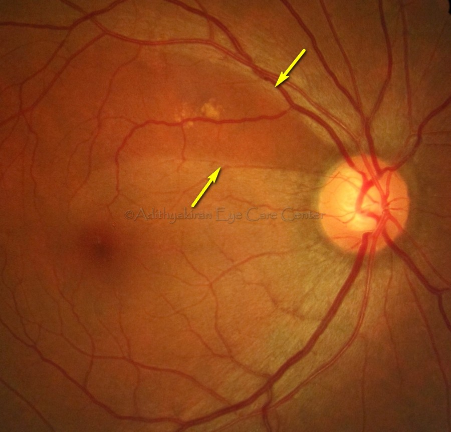
This ‘cracking’ is called lattice degeneration (see above). These cracks can eventually stretch too much to form ‘tears’ or ‘holes’. (see below)

vodvos-retinal-hole

Once a hole or tear appears you might get symptoms of streaks of light flashing past your face. These flashes can be normal too. The next time you feel a sneeze coming up just close your eyes and let go. You’ll see white spots of light flying on the periphery of your vision that will last for a few seconds.

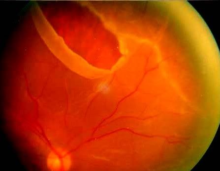
Sometimes, especially in higher degrees of myopia the vitreous (the jelly like substance inside the eye) liquifies and it is this liquified vitreous that appears as small dots or shadows (called floaters) in your vision when you look at a bright background. If this fluid vitreous goes though the tear it can separate the retina from the underlying layers, a condition called retinal detachment. (see below)

RD

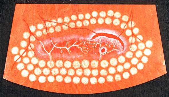
There will be a sudden drop in vision with flashes that are continuous. This is an ophthalmic emergency. Unless the retina is surgically replaced immediately there is great risk of permanent loss of vision. Retinal detachment can be prevented to a large extent by using lasers to ‘fence off’ the hole (see below) and prevent the liquid vitreous going under the retina.

lasered

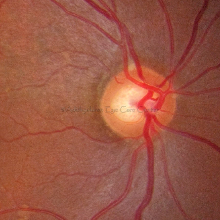
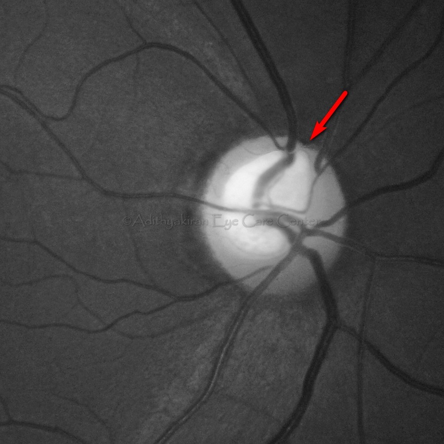
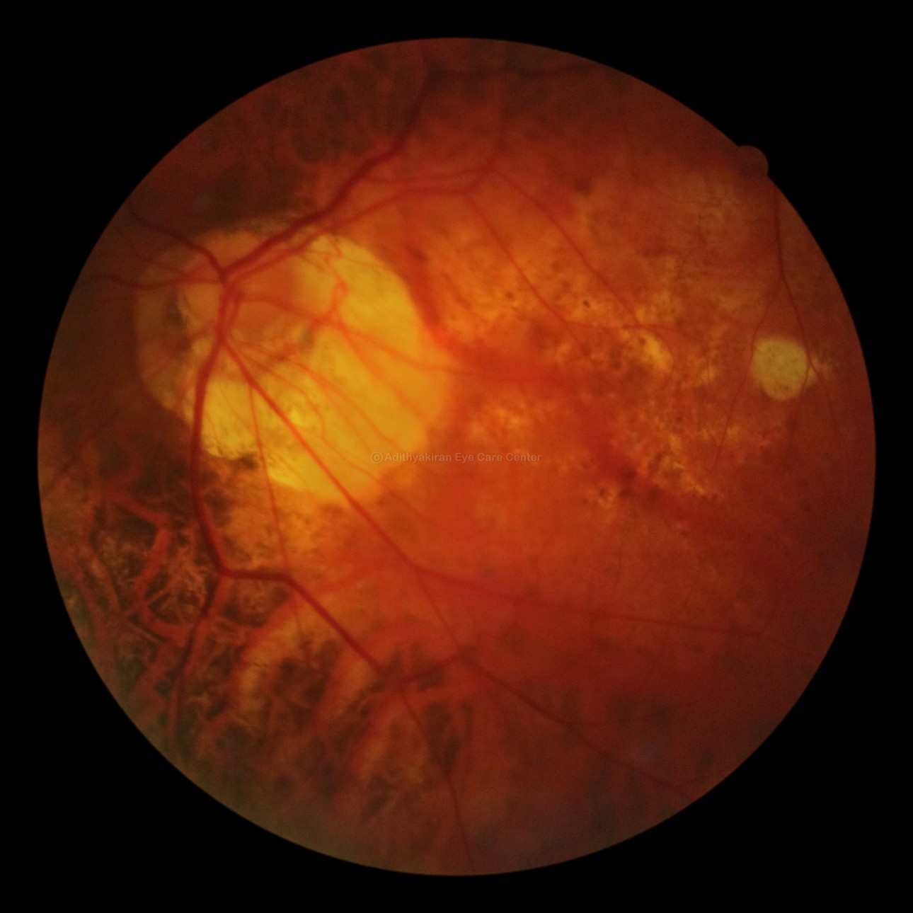
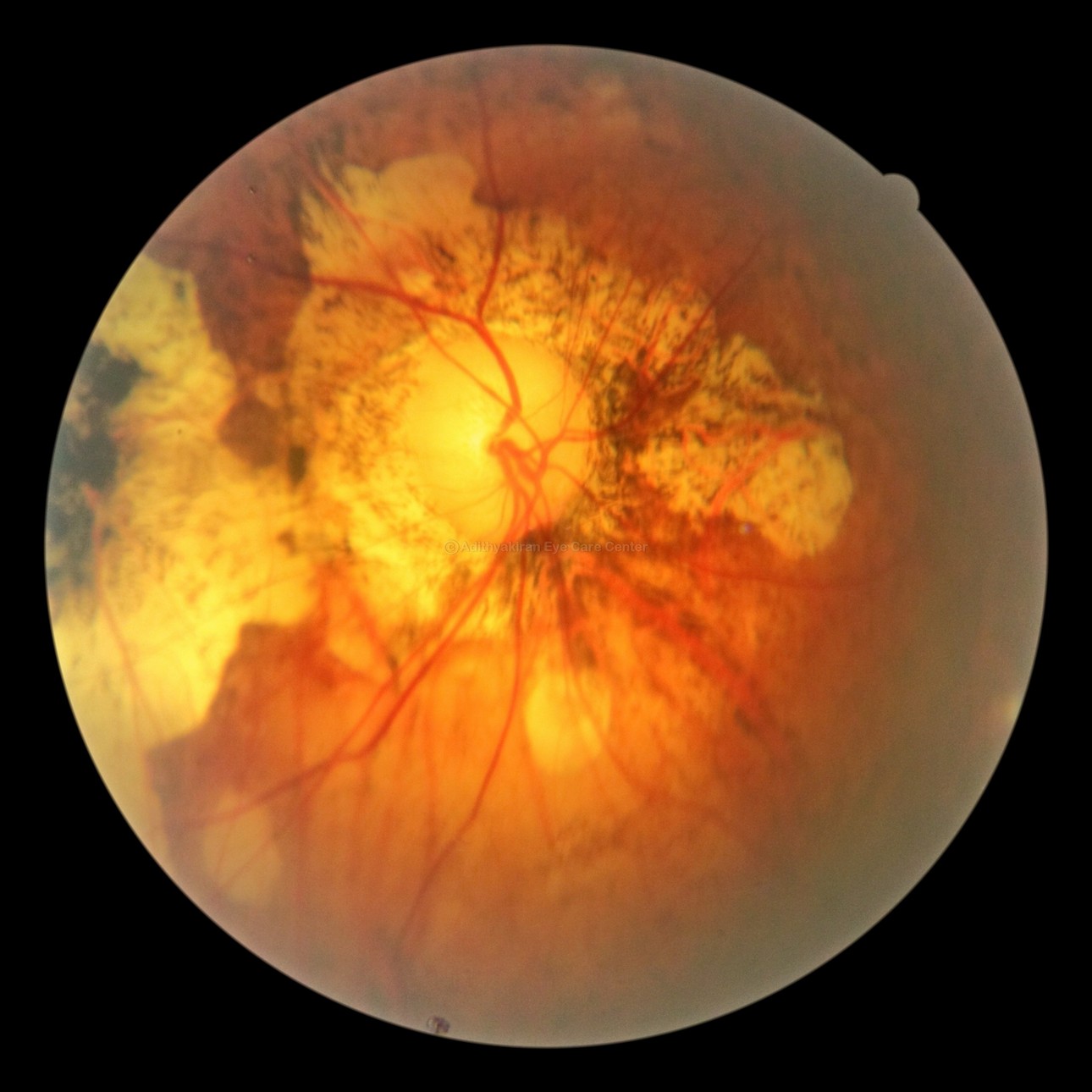
In some advanced cases of myopia the eyeball stretches backwards so much that the retina and underlying structures get thinned and damaged near the optic nerve head and the macula (the part of the eye that controls fine vision). That condition is called a posterior staphyloma and it usually results in poor central vision.

early staphyloma Pathological myopia

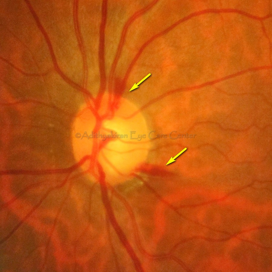
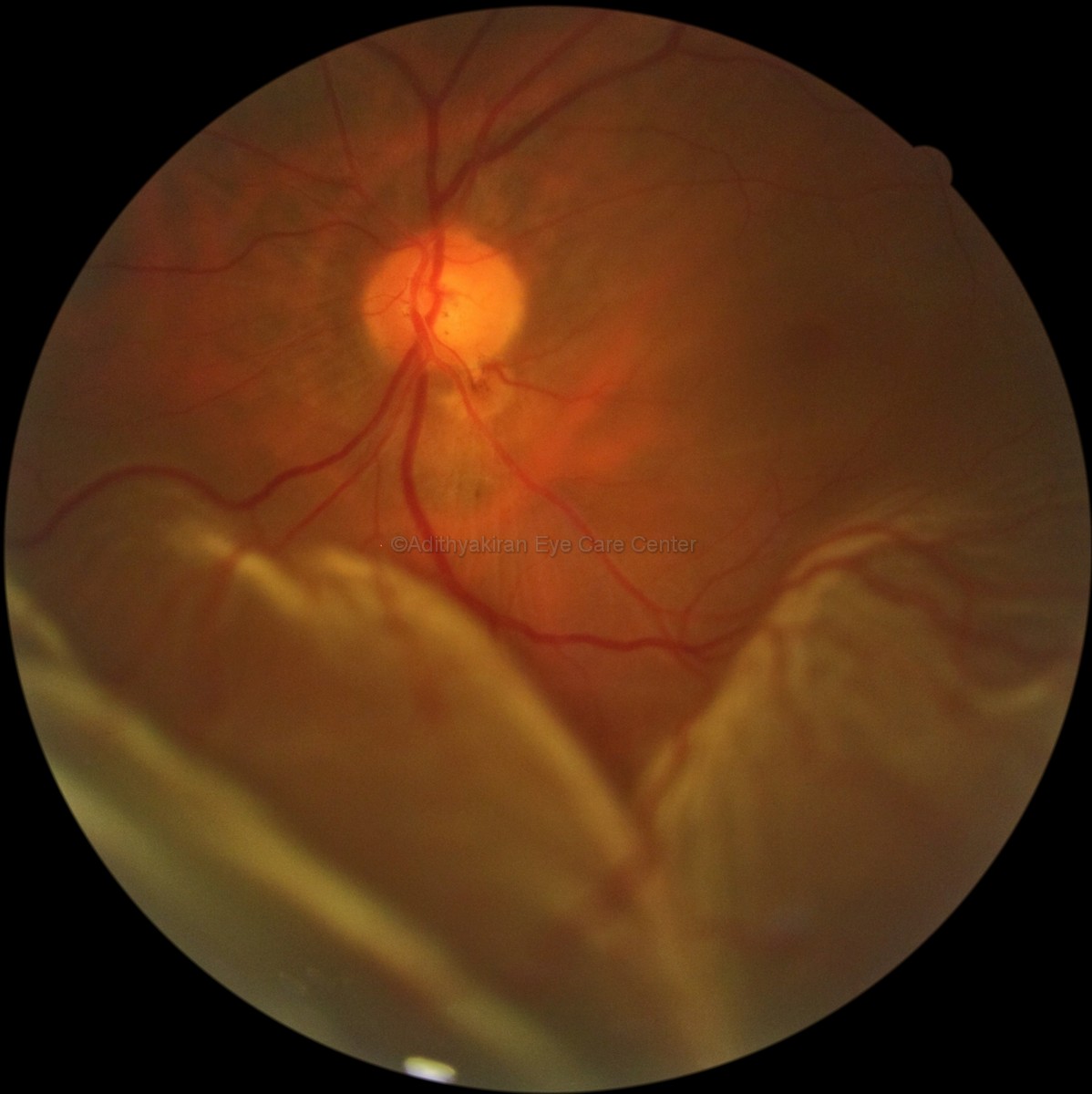
Compare this to a relatively normal fundus in this picture below.The disc and the blood vessels are spread out on a very uniform background retina unlike in the pictures above.

normal fundus

So the next time you go to your ophthalmologist don’t just say, “I’ve come to check my number”; say “I’m here to check my eyes inside and out”.

Make sure you get all these tests done:

  1. Visual acuity with and without spectacles
  2. Best Corrected Visual Acuity if your spectacles feel inadequate
  3. Intraocular pressure
  4. Retiniscopy
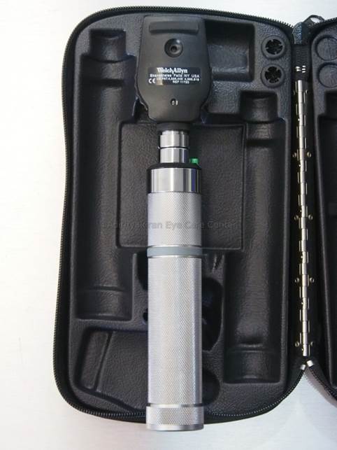
  5. Ophthalmoscopy, including indirect ophthalmoscopy if you have higher degrees of myopia

EVENING UPDATE 6.30PM

I’m updating here because of a patient who walked in an hour ago. She had undergone LASIK in 2002 and her spectacle number before the procedure was -14 diopters. After LASIK it had reached near about zero and is still stable. If you have undergone LASIK to get rid of your glasses please do keep in mind that this procedure only alters the front of your eyes. All the serious changes occur within the eye and therefore you need to continue getting your eyes checked every year, as described above. Otherwise, subtle progress can be missed and you could end up with a sight threatening complication!

 
2 Comments

Posted by on June 20, 2013 in Uncategorized


Tags: , , , , , , , , , , , , ,

Glaucoma (Part 3) – “Confirming the presence of the Silent Thief”

In the previous part I had told you how to identify if you are a potential candidate for developing Glaucoma, the Silent Thief of sight. Now it is time to look at the different ways you can confirm if you have the disease.

Detecting glaucoma, in most instances, does not require sophisticated equipment. Ophthalmologists can identify features of the disease by just using some common instruments that they already possess. If a systematic approach is done the disease can be picked up early.

1. SLIT LAMP EXAMINATION

Slit Lamp

During preliminary examination with the instrument called the slit lamp the ophthalmologist may see some features that point to the presence of glaucoma. There maybe shallowing of the anterior chamber, the space between the cornea and the iris suggesting a possibility of angle closure glaucoma. There can be pigments on the inner surface of the cornea, or some flaky white material on the surface of the lens called pseudo-exfoliation.

2. TONOMETRY

The next step will be to see if your intraocular pressure (IOP) is raised. Remember though, glaucoma needn’t always be associated with an elevated IOP. There are eyes that have glaucoma even with normal or low pressures. IOP, therefore, is not the main criteria for deciding if you have glaucoma. IOP is measured by an instrument called the applanation tonometer, attached to the slit lamp. After instilling a drop of anesthetic the tip of the instrument is made to touch the dome of your cornea and a reading is taken from a rotating dial on the side of the instrument. There are other instruments to measure the IOP but the applanation tonometer is take as standard.

GAT

3.PACHYMETRY

Sometimes the IOP maybe normal or below normal but other features may give an indication that the eye has been affected significantly by glaucoma. The ophthalmologist may then do a pachymetry, a test done to measure the thickness of the cornea.

Pachymeter


It is not unusual to have people with myopia (short sightedness) to have a thin walled eye. This gives a falsely low reading of IOP. The reverse maybe true in other eyes. Due to a thicker cornea, the recorded IOP might by high, but there may not be any evidence to suggest glaucoma. Such eyes with higher IOP but without features of glaucoma are designated as Ocular Hypertension, and will need follow up to see if they progress to glaucoma.

4. OPTIC NERVE HEAD EVALUATION

After having measured the IOP the next step is to look inside your eye to see if there is any physical damage to your optic nerve head (ONH). This is the most important part of the examination that lets the ophthalmologist decide if your eye has been affected by glaucoma and if you need any treatment. The ONH can be examined by a simple hand held instrument called the ophthalmoscope; a self illuminating, hand held instrument that is held close to your eye and the doctor looks inside from the other side.

ophthalmoscope

For greater details, a small, powerful hand held lens (a 78 or 90 diopter) is used in combination with the slit lamp bio-microscope. It gives a bigger 3D view of the ONH and any damage can be assessed easily.

90D lens

This is the most important step in the assessment of a person suspected to have glaucoma. Features onthe ONH or surrounding retina can instantly clinch the diagnosis of glaucoma. The doctor will look for certain changes in the ONH that will show if glaucoma has already damaged the eye. I will dwell a little longer on this part of the exemination because, of all the examinations, this is the single most critical test that gives the doctor a hint that you have glaucoma.

Normal optic disc

To know what features suggest damage to the doctor, you should know what a normal ONH looks like. The optic nerve is the bundle of nerve fibres that start from your retina and go towards the visual cortex of your brain. As they exit the eye they form a distinct round or slightly oval disc, orange in colour, called the optic disc (see figure above). In the center of the disc (blue circle in figure below) is a smaller, light yellow coloured portion called the optic cup (yellow circle in figure below). This cup is usually well centered or slightly eccentric, when surrounded by heathly nerve fibers.

Disc & cup


The portion between the blue and yellow ring represents healthy nerve fibers and they are present in all quadrants of the disc in slightly varying widths. This part is called the neuro-retinal rim (NRR). The first sign of damage is a narrowing of the NRR, (red arrow) sometimes all the way upto the edge of the disc.

Rim narrowing

Sometimes associated with this narrowed rim there can be other signs that indicate the presence of active glaucoma. There might be small hemorrhages (bleeding) along the disc margin (yellow arrows in the picture below).

Hemorrhages

In some eyes, there is a dark band on the retina corresponding to the area of narrowing of NRR (between yellow arrows in the picture below). They are areas of retinal thinning due to loss of some tissue there. They are called retinal nerve fiber layer (RNFL) defects. They usually correspond to defects in the visual field tests.

RNFL defect

You can see that an ophthalmologist can make an instantaneous diagnosis of glaucoma if any of the above mentioned findings are there in a patient’s eye. Very simple instruments and equipment are need to make a diagnosis. It is only in cases where the findings are borderline that we need sophisticated instruments to give additional information to support the diagnosis.

Once glaucoma is detected the next step is to assess how much damage it has done to the eye. Damage to visual function usually occurs from the periphery of your vision. This is assessed by a test that plots the visual field. More about that in the next post. 

 
Leave a comment

Posted by on May 30, 2013 in Eye health


Tags: , , , , , , , , , , , , , , ,